


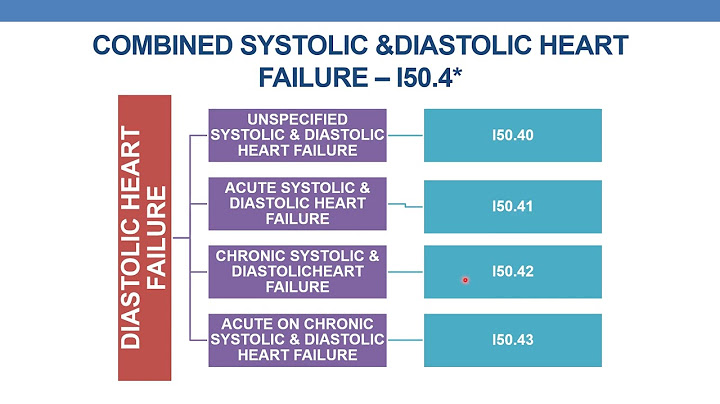
How to Cite This Chapter: Ibrahim O, Jaeschke R, Szczeklik W, Jankowski M. Jugular Venous Distention. McMaster Textbook of Internal Medicine. Kraków: Medycyna Praktyczna. https://empendium.com/mcmtextbook/chapter/B31.I.1.120. Accessed October 11, 2022. Show Last Updated: July 7, 2020 Last Reviewed: April 10, 2021 Chapter Information McMaster University Editorial Office Section Editors: Roman Jaeschke Authors: Omar Ibrahim, Roman Jaeschke Polish Institute for Evidence Based Medicine Editorial Office Section Editors: Andrzej Budaj, Wiktoria Leśniak Authors: Wojciech Szczeklik, Miłosz Jankowski Main Documents Taken Into Account: Chua Chiaco JM, Parikh NI, Fergusson DJ. The jugular venous pressure revisited. Cleve Clin J Med. 2013 Oct;80(10):638-44. doi: 10.3949/ccjm.80a.13039. PMID: 24085809; PMCID: PMC4865399. Vinayak AG, Levitt J, Gehlbach B, Pohlman AS, Hall JB, Kress JP. Usefulness of the external jugular vein examination in detecting abnormal central venous pressure in critically ill patients. Arch Intern Med. 2006 Oct 23;166(19):2132-7. doi: 10.1001/archinte.166.19.2132. PMID: 17060544. Seth R, Magner P, Matzinger F, van Walraven C. How far is the sternal angle from the mid-right atrium? J Gen Intern Med. 2002 Nov;17(11):852-6. doi: 10.1046/j.1525-1497.2002.20101.x. PMID: 12406357; PMCID: PMC1495124. Cook DJ, Simel DL. The Rational Clinical Examination. Does this patient have abnormal central venous pressure? JAMA. 1996 Feb 28;275(8):630-4. PMID: 8594245. DefinitionTop Jugular venous pressure (JVP) was once among the most central parts of the physical examination. Widespread use of numerous imaging techniques has diminished its importance; nevertheless, it provides important diagnostic information regarding volume status and cardiac pressures. JVP is assessed clinically by positioning the patient supine with the head of the bed elevated between 30 and 45 degrees. It is visualized as a multiphasic pulse between the 2 heads of the sternocleidomastoid muscle, and if not immediately visible, it can be visualized by elevating or lowering the head of the bed until it comes into view. The height of this pulsation is then measured relative to the sternal angle. The sternal angle is ~5 cm above the right atrium independent of position; however, this may be an underestimate, particularly in older patients as well as in those with obesity or hyperinflation. JVP height above the sternal angle can be used to estimate the right atrial pressure. A JVP of 0 to 4 cm above the sternal angle is considered normal, whereas a JVP >4 cm is considered jugular venous distension. Compared with the carotid pulse, the internal jugular vein pulse is multiphasic, nonpalpable, occludable with digital pressure, rises as the angle of the bed is lowered, falls as the angle of the bed is raised, falls with inspiration, raises with expiration, increases with abdominal pressure, is lateral in position, and is predominantly inward (downstroke). Certain abnormalities in the character of the JVP are associated with specific cardiovascular abnormalities. Applying pressure over the upper abdomen can increase venous pressure via the abdominojugular reflux (AJR). If the JVP elevates >3 cm for >10 to 15 seconds following application of abdominal pressure, this finding is specific for elevated central venous pressure. Notably, obstructive pathology affecting the vena cava may cause abdominal pressure not to transmit freely to the jugular veins, causing an absent or diminished AJR. Alternatively, filling of the external jugular veins may be assessed in some circumstances. Gross distension of the external jugular veins is often more easily observed than distension of the deeper internal jugular vein but it is less specific. Causes and PathogenesisTop Venous distention is due to an increase in the venous blood pressure. Causes of jugular venous distention include: 1) Bilateral distention: Right (with or without left) ventricular failure, large pericardial effusion (including cardiac tamponade), constrictive pericarditis (often with paradoxical increase in JVP during inspiration, known as Kussmaul sign), restrictive cardiomyopathy, tension pneumothorax, mechanical obstruction of the superior vena cava (eg, by thoracic tumors, mediastinal lymphadenopathy, thrombosis, fibrosis, or aneurysms), superior vena cava syndrome (see Superior Vena Cava Syndrome), tricuspid stenosis or regurgitation (tricuspid regurgitation results in c-v waves with systolic increase in the venous pulse), pulmonary hypertension, pulmonary embolism. 2) Unilateral distention: Localized mechanical obstruction due to, for example, tumors (including goiters), thrombosis, large aneurysms, or lymphadenopathy. Left-sided distention may be caused by compression of the left brachiocephalic vein by an aortic aneurysm. DiagnosisTop 1. Assess the vital signs (breathing, pulse, blood pressure [BP]), as the underlying condition may be life threatening (particularly cardiac tamponade, acute heart failure, tension pneumothorax, or pulmonary embolism). 2. Take a history and perform a cardiovascular examination: Elements of the past medical history, clinical context, associated symptoms, and timelines often help clarify the differential diagnosis. Findings of cardiovascular and respiratory examinations are associated with specific diagnoses. The AJR may help localize the cause. A pulsus paradoxus (difference in systolic BP between expiration and inspiration >10 mm Hg) should be examined if concerned for tamponade or obstructive lung disease. This is classically not present in restrictive cardiomyopathy or constrictive pericarditis, however, Kussmaul sign (an elevation of the JVP with inspiration) is present in these conditions. 3. Potential other diagnostic studies: Chest radiography, echocardiography, or both in patients with suspected heart failure, cardiac tamponade, constrictive pericarditis, or valvular heart disease. Ultrasonography of the neck as well as serum thyroid-stimulating hormone (TSH) and thyroid hormone levels in patients with a large goiter. Computed tomography (CT) of the chest, neck, or both in patients with superior vena cava syndrome or suspicion of cancer, lymphadenopathy, fibrosis, or aneurysm (accompanied by edema of the face and neck and distention of the veins of the upper half of the chest). Bronchoscopy as directed by other findings. Computed tomography angiography (CTA) in patients with suspected pulmonary embolism, intrathoracic tumor, or a large aneurysm. What causes left jugular vein distention?JVD is a condition wherein increased blood pressure causes your jugular vein to bulge. Many cardiovascular conditions — including heart failure, high blood pressure, and fluid accumulation around the heart — can cause JVD.
Do you have distended neck veins with rightFor people with right-sided heart failure, the left side of the heart has usually already failed, so the right side is under much more pressure to pump blood. Over time, the right side is weakened and cannot work as well. Blood then accumulates in the veins and leads to a bulging jugular vein.
Why does JVP increase in left heart failure?Elevated jugular venous pressure is a manifestation of abnormal right heart dynamics, mostly commonly reflecting elevated pulmonary capillary wedge pressure from left heart failure. This usually implies fluid overload, indicating the need for diuresis.
Is JVP raised in heart failure?Elevated jugular venous pressure reflects increased right atrial pressure, which itself correlates with elevated left-sided filling pressures in patients with chronic heart failure.
|

Left sided heart failure symptoms jugular vein distention
Related Posts


Copyright © 2024 berikutyang Inc.